


News
Startup RoboScope automates pathomorphological examinations in Russia
In order to confirm the presence of a tumor in a person, classify it and select the optimal treatment, it is necessary to conduct a pathomorphological study. More than 7 million such analyzes are performed annually in Russia, most of them using the most common analog microscope. Startup RoboScope intends to change this, thereby increasing the speed, accuracy and quality of research. The editors of INNOVATIONS talked to the CEO of the project, Ilya Efremov.
— How was the idea of creating RoboScope born?
“It all started with a dry drop of urine. The scientific director of our project, Igor Shaderkin, is a urologist, and he needed a microscope to assess lithogenic (stone-forming) activity in patients with urolithiasis from a dry drop of urine. Our team decided to initiate the creation of an automated microscope to digitize the microscopic picture of a dried urine drop. Subsequently, this microscope went through several stages of modernization, and at one of the stages we realized that it could also be used in pathomorphology.
In the process of work, we began to be interested in what equipment pathomorphological laboratories use, to study the market. From the 2020 statistical study “The State and Main Tasks of the Pathological Anatomical Service of the Russian Federation”, we learned that over 7 million pathomorphological studies are performed in Russia a year. To do this, experts must look under a microscope for about 42 million glasses. The workload is constantly growing, and the staff of the service is understaffed.
The entire research process - from the preparation of histological preparations (they are smears, organ prints, film preparations, thin sections of pieces of organs stained with one or another dye) to directly performing microscopy, is usually performed in the old fashioned way, manually. Almost 40% of the pathomorphological equipment fleet in the country is over ten years old and needs to be replaced.
From time to time, situations arise when doctors need to double-check something, compare samples in order to understand how the disease developed, or even prove the correctness of their conclusions in the event of a lawsuit, and the “glass” with the histological preparation is no longer suitable for use. Its shelf life is limited.
Ilya Efremov
Finally, in many medical institutions, clinicians do not have online access to pathological examination results, either because such a technical possibility is not available or they use the services of a third-party laboratory. This means that the results of the analysis have to wait quite a long time. They can get lost, they can be transferred to the wrong organization, to the wrong doctor, and this is also a problem.
Thus, we saw the demand to automate the entire research cycle. Of course, we were faced with the task of making our solutions as accessible as possible. After all, today on the market there are means of automating the pathomorphological service produced abroad, but they are very expensive.
- What numbers are we talking about?
- We try to keep within the limits of 2 million rubles. For comparison, similar imported complexes cost about 10 million and more.
What does the buyer get for this money?
“One of the central ideas of our project is to save the doctor from having to be near the microscope. Glasses can be loaded into the installation by paramedical personnel, a laboratory assistant. The doctor will be able to view already digitized images in his office or remotely from anywhere in the world.
— What technology underlies your product and how justified is the prefix “robo” in its name?
- We use WSI (Whole Slide Imaging) - a technology for stitching multiple fields of view into one large one, which is the basis of scanning. That is, small fragments are digitized, and then the program stitches them into one large canvas. The prefix in the name RoboScope MARK-1 implies that it is a three-axis robot.
- What does your product look like?
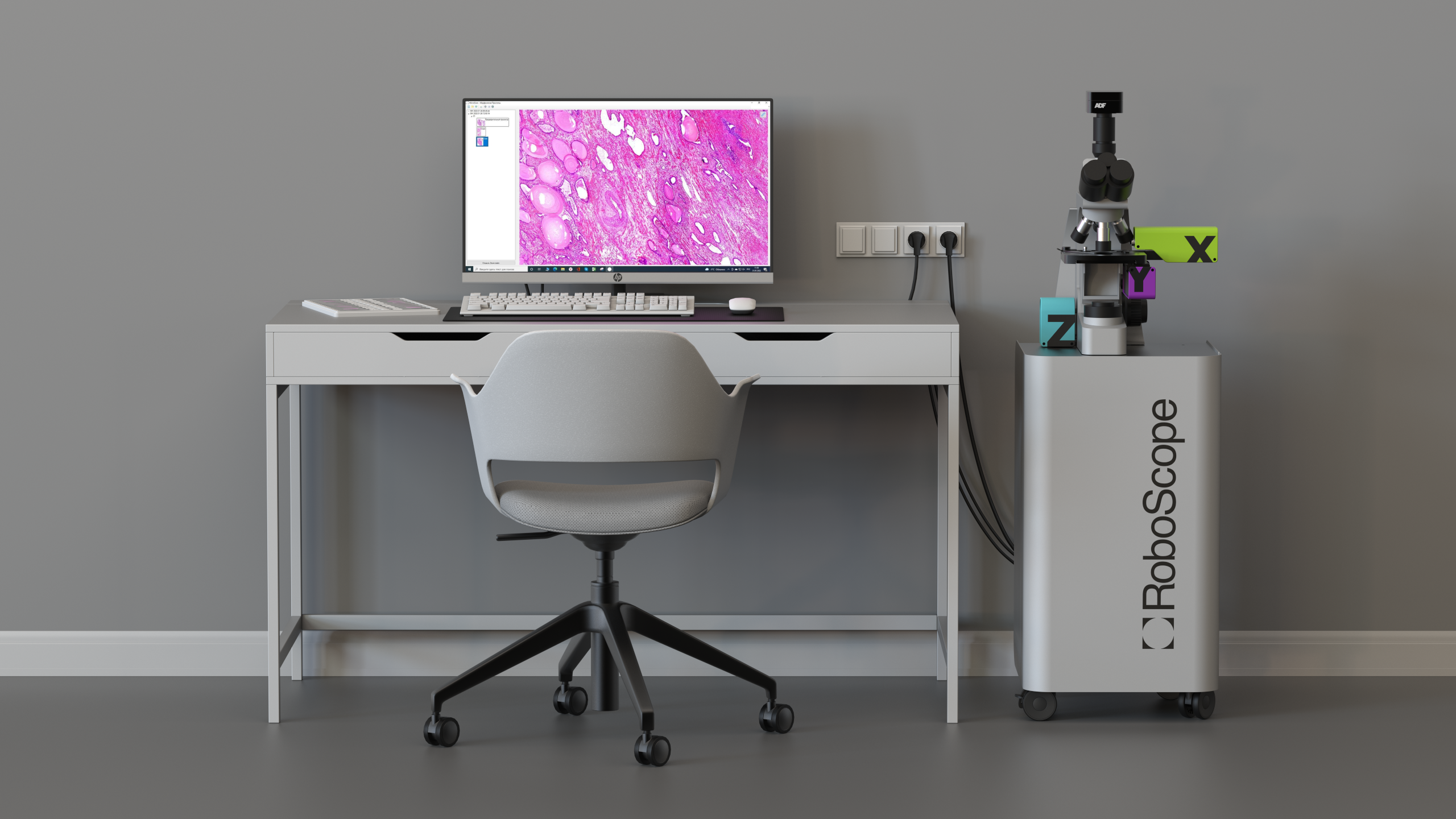
“The complex consists of a control and data analysis system that looks like a cabinet, a light microscope with a movable table and a camera. RoboScope will be delivered with a monitor and a mouse right away, which means that the specialist will receive a ready-made solution. He will not need to buy a computer, for example, in order to start working with our complex. No need to somehow prepare the room for the installation of RoboScope. And its use does not require special training.
From the point of view of hardware, there is nothing difficult for a laboratory assistant who will use our equipment. Minimal light microscope skills are required to correctly set the light in order to obtain a high-quality image, in order to change the lens and histological specimen if necessary. These are fairly simple steps that most healthcare professionals are familiar with.
As for the software, we will soon complete the redesign of our program so that it is intuitive. In any case, the scanning and digitization of glasses takes place in the open and widely used DICOM format (Digital Imaging and Communications in Medicine) in Russia and the world. This is an industry standard for storing and distributing medical images. In addition to the picture itself, each file contains information about the patient: his name, age, as well as the number of the image, and the designations of the scanning modes.
The DICOM format is supported by most medical device manufacturers and software developers for healthcare organizations. And our program will have a standard set of tools for working with DICOM data: ruler, zoom, preview, etc. Thanks to this, the complex is easily integrated: it is convenient to store the accumulated data, upload it to any medical information system (MIS), laboratory information system (LIS) or telemedicine platform.
In the future, we will be able to create datasets and, using machine learning and artificial intelligence, create content for training on virtual histological preparations, and develop telementoring. All this will contribute to improving the quality of training of pathomorphological service specialists.
— Who might be interested in your development, except for pathomorphological departments?
— There is interest in our product from both the medical and scientific communities. For example, we had several requests from doctors who saw our presentation at industry forums. They asked when RoboScope would be ready and when it would be available for purchase. It can also be requested in forensic and reference centers (which issue an expert "second" opinion on complex clinical cases).
— At what expense is the project being implemented?
- So far, these are the own investments of the startup participants. In total, we have invested 120 thousand dollars, or 12 million rubles. Recently, we received the status of a Skolkovo resident and are counting on grants from this fund. They also applied for a grant from the Foundation for Assistance to the Development of Small Forms of Enterprises in the Scientific and Technical Field of Bortnik. We are negotiating with strategic partners to attract investment.
— What is your business model?
We are considering different options. Understanding that it is difficult for medical organizations to pay a large amount for equipment one-time, we are ready to cooperate with them as part of a subscription, according to the cost-sharing scheme, in which equipment is provided free of charge, and monetization occurs by charging a fee for research (for glass). We are going to offer service with a permanent upgrade aimed at expanding the functionality of the complex.
- Who is on your team?
— In addition to me, the technical director and engineers, there is a medical expert in our team: MD. Professor of the Department of Pathological Anatomy named after A.I. Strukov First Moscow State Medical University. THEM. Sechenov, doctor of the highest qualification category in pathological anatomy Alexander Tertychny and scientific adviser Ph.D., head of the laboratory of the Institute of Digital Medicine of the First Moscow State Medical University. THEM. Sechenov Igor Shaderkin.
- How much do you depend on foreign components and at what production facilities do you plan to produce the complex?
— Our product has some imported components, but they are all produced in the Asian region. We are completely independent from European and American suppliers. As for the software part, it is either own development or open source.
At the moment, we have an agreement with the Delrus company on the use of their capacities for the production of our product.
- At what stage is the project now?
- The MVP of the complex called RoboScope MARK-1 is ready. Now we are preparing to register it with Roszdravnadzor as a medical device.
But the light microscope is not our only goal. We position ourselves as an RnD company, so we are going to develop further in this direction.
Our next product will be the same complex, but a class higher than RoboScope MARK-2. Then we will make robots for sample preparation of pathomorphology: dehydration of histological preparations and staining of histological preparations. We want to fully automate the entire cycle of work of the pathomorphological service.
Roboscope Patology LLC
Политика конфиденциальности
Согласие на обработку персональных данных
Moscow

2025